Nashwan Saleh Mohammed Al-ashwal
Al-Nasr general hospital and Al-ashwal clinic for medical & cardiac diseases || Adhala || Yemen
DOI PDFIntroduction:
In fact and as we know medically that coronary arteries diseases causes typical chest pain as retrosternal pain that radiating to right and left shoulder , with positive electrocardiogram changes but can be seen with normal or non-specific traces in spite of patient’s complaint [1], which necessitate the demand for easily accessible means to view the coronary arteries atherosclerosis, like the transthoracic echocardiography , eventually, by it we can detect the earliest signs of myocardial blood insufficiency prior to infarctions and heart failure.
Presentation:
A patient of 40 years old male, married and father for 6 children, he had been actively working in his farm, and he was complaining of chest tightness and back pain since January2017 with progressive manner, the pain and tightness were persisted for 5-10 minutes After he had walked up hill then it was subsided gradually with no medical interventions, the second attack was occurred 5 months later after nervous occasions and had persisted for 30 minutes with no associated sweating , his wife was performed a chest massage that relieving the pain, but his maneuver was failed in the third attack at the end of July 2017 after he had climbed the house stair then he had been looked for a medical advice in many specialized clinics of chest and cardiac diseases where he was informed about his elevated blood pressure of 160/98mmhg and his investigations of electrocardiogram and echocardiogram were normal with relative disturbances of low density and high density lipoproteins value with normal total cholesterol and triglycerides. SO, the doctors prescribed beta-blocker (bisoprolol 5mg/per day), anxiolytic and proton pump inhibitor’s drugs for him , but after 2 months of treatments while he was using his drugs, he came to my clinic with a complaint of chest tightness and back pain that intermittently increased and decreased since 3 days and he had taken drugs for fibromyalgia (ketoprofen 25 mg every day and baclofen 10mg two times a day) as prescribed by doctors but without benefits.
No nausea, vomiting, acid or food regurgitations, sweating, headache or wheezing.
He was suffering of hyperacidity and epigastric pain as he was diagnosed of helicobacter pylori gastritis with positive h. Pylori IgG two years ago.
No diabetes, hypertension, ischemic heart disease or tumor history in his own family.
Investigations: of blood has normal results of blood count , sugar and electrolytes level. Increase in lactate dehydrogenase 600 u/l, increase in low density lipoproteins 140mg/dl, decreasing in high density lipoproteins 40mg/dl with normal cholesterol, triglycerides, troponin I and creatinine kinase-MB.
Physical examinations: Revealed elevated blood pressure 160/98mmhg, in setting position with normal radial pulse rate of 90 beats per minute with good volume and regularity, normal heart and respiratory sounds, distended abdomen with mild bilateral subcostal tenderness more to the left, tympanic percussion left para umbilical region, no tenderness over the costal ribs or the vertebral bones.
Electrocardiogram: (heart tracing): Revealed a non-specific changes, for example, (low amplitude of III, and aVL limb leads with notching of QRS complex in previous limbs leads and V1 of chest leads) as shown in photo( 1).
Echocardiography: (heart imaging by ultrasound): Reveals a normal cardiac chambers dimensions, valves and systolic fractional contractions(ejection fraction=53% & fractional shortening =27%) without resting wall motion abnormalities, but long axes of inferior and posterior walls is less than the lateral axis amplitudes, as shown in (photo2,3 and 4). And the coronary arteries walls are thickened with irregular internal lines and decreased lumens diameters, such as main right, posterior descending and left circumflex walls thickening as shown in photo (6) with thickened wall and focal atheroma in the left anterior descending coronary artery as shown in (photo 5 ). So , what am trying to say that the Diagnosis is coronary arteries atherosclerosis with variable stenosis of the right main, posterior descending and left circumflex coronary arteries with preserved cardiac chambers dimensions and functions at rest.
Treating by two steps: immediately, he had given sublingual isosorbid dinitrate 5 mg so. his complaint was subsided after 3 minutes and it returns after 10 minutes again. for that, intravenous injections of one solcoseryl ampoule (20mg hemodialysate), actovegin ampoule 200 mg and salicylic acid vials 900 mg were given to increase the tissues oxygen, dilating arteries and preventing platelets aggregation respectively then the patient felt better and be able to take a comfortable breath.
Home therapy was as follows: antiplatelet aggregation clopidogrel 75 mg twice a day for 3 days then one tablet daily, tri omega 500 mg twice a day and solcoseryl 20 mg injected intravenously once a day for 5 days with follow up for 18 months.
Follow up: The period from the first 10 days up to three months blood pressure level decreased to 145/85 mmhg , no chest tightness or back pain, and by transthoracic echocardiography there are increasing of contraction activity as ejection fraction to 65% and increasing in long axes amplitudes of posterior wall and inferior wall as shown in photo (7 & 8 )., also after nine months there is no recorded complaints and no changes in low or high density lipoproteins levels but there was improving in blood pressure level to 135/80 mmhg without giving him any anti-hypertensive drugs.
Results: Firstly, The three long axes amplitude values deteriorated below 10 mm for posterior and inferior axes and below 20 mm for right ventricle axis were compared with 3 normal persons values that are more than 10 mm up to 17 mm for left ventricle axes and more than 20 mm for right ventricle axis except for one normal person whose axis value of posterior wall was 10.5 mm in lower level than his own other axes, and also it was compared with values of one patient’s with ischemic cardiomyopathy since one year diagnosed, and his values was below 8 mm for left ventricle axes and below 12 mm for right ventricle axis, as shown in tables (1 ) and figures(1),before therapy,
Table (1) represent the cardiac long axes amplitudes values of the presented patient before therapy, 3 normal persons and patient of ischemic cardiomyopathy .
| cases | Measurements in mm*
Lateral septal anterior inferior posterior RV† |
|||||
| Patient 1 | 13.3 | 14.5 | 13.5 | 6.5 | 4.7 | 17.8 |
| Normal 1 | 16.5 | 17.7 | 16.5 | 16.5 | 13.5 | 22.5 |
| Normal 2 | 13.3 | 16.1 | 13.3 | 13.3 | 10.5 | 22.2 |
| Normal 3 | 14.7 | 14.7 | 15.5 | 14.5 | 14.5 | 19.7 |
| Patient 2 | 6.4 | 6.4 | 6.4 | 7.0 | 6.4 | 11.7 |
mm*= millimeters. RV† = right ventricle free wall axis.
FIGURE (1) Represent the walls long axes amplitudes for the patient , 3 normal persons and another patient , it reveals a decrease in two axis below 10 mm of the presented patient (1) walls axes and decreasing in the all axes of the patient (2) with ischemic cardiomyopathy.
also it was compared with the deteriorated axes of the patient’s with ischemic dilated cardiomyopathy revealing improvements in the deteriorated and also the normal axes values of the presented patient after therapy, as shown in table (2) and figure (2). Secondly, the coronary arteries , were visibly seen by using the short axis view
Table (2) represent the cardiac long axes amplitudes values of the presented patient and patient of ischemic cardiomyopathy after therapy.
| cases | Measurements in mm*
Lateral septal anterior inferior posterior RV† |
|||||
| Patient 1 | 15.6 | 15.5 | 14.9 | 9.2 | 7.8 | 20.5 |
| Patient 2 | 6.4 | 6.4 | 6.4 | 7.0 | 6.4 | 11.7 |
*mm= millimeters. † RV = right ventricle free wall axis.
FIGURE (2) Represent compare between the presented patient long axes amplitudes values with the ischemic dilated cardiomyopathy values and the treatment response by increasing axes amplitudes of the presented patient, reflect the importance of early diagnosis to avoid permanent myocardial damage.
, during echocardiographic examination just at level of aortic valve with angulation toward the right shoulder for right one, and mildly toward the left nipple for both left main stem or proximal left anterior descending and more angulation for left circumflex in separated arterial presentation variant as shown in photo (6), and part of the mid level of left anterior descending seen by long parasternal view with slight upward angulation while the posterior descending one seen by short axis view as shown in photo (5), and the ability to measure the parts of the visualized coronary arteries walls and lumens to detect the narrowed sites as shown in table (3) in which I have putting the lower and upper normal diameters depending on distance compare between the same arteries points in the same patient’s and other normal persons coronary arteries diameters.
Table (3) represent coronary arteries walls and lumens diameters by transthoracic echocardiography.
| Coronary arteries | Measuring in mm*
Wall thickness lumen patency |
|
| Main right stem | 0.7-1.2 | 2.0-2.9 |
| Posterior descending | 0.5-0.8 | 1.5-1.9 |
| Left main stem | 0.9-1.15 | 2.2-3.2 |
| Anterior descending | 0.65-0.97 | 1.6-2.1 |
| Left circumflex | 0.55-0.76 | 1.1-1.5 |
*mm= measuring unit of distance millimeters.
Discussion:
At present many medical equipment are used for diagnosis of chest discomforts that ranging from simple chest pain or tightness to severe fatal pain, for saving human lives, for example, electrocardiography that tracing the electrical activities of the heart in form of systolic and diastolic waves and the echocardiography that imaging the heart activities and showing its chambers, valves and walls motion, and this thing leads to improve in patient’s health, except for some percentage of limitations in which the pathological signs weren’t clear or shown in the early stage of the disease such as in electrocardiography, as said: Kevin Channer et al. A trace can suggest, for example, that a patient’s heart is entirely normal while in fact he or she has severe and widespread coronary artery disease. In addition, some of these patients are less than half of patients presenting to hospital with an acute myocardial infarction will have the typical and diagnostic electrocardiographic changes present on their initial trace, and as many as 20% of patients will have a normal or near normal electrocardiogram [2], Even by exercise stress test , as Fedorowski & Redden said: there is a false positive and false negative results in exercise stress test results depending on the high or low pre-test probability respectively[3], and as in this case, the patient has suffering but has non- specific electrocardiogram changes in leads III, aVL and V1 QRS with normal echocardiogram in name of chambers, valves and ventricular functions with ejection fraction of 53%, and its manifestations not improved by beta-blocker 5mg, anxiolytic and acid release inhibitor’s but improvement began with given him the sublingual isosorbid dinitrate 5 mg and establishing it by the solcoseryl, actovegin and salicylic acid 900 mg injectable drugs, which enhancing the diagnosis of coronary arteries atherosclerosis and emphasizing it, therefore, while many patients had been suffering of the same manifestations and were underwent exercise stress test but their results were normal as in echocardiogram , regrettably” some of them developed heart failure one year later with ejection fraction of between 40-48% and one’s die hard after one month due to massive myocardial infarction as a result of misleading diagnosis in the absence of supporting investigations indicating to the presence of underlying pathological process involving the coronary arteries, this additionally, necessitates the need of the presence of easy accessible, accurate and useful equipment like transthoracic echocardiography, therefore its applying to this patient reveals its importance and abilities to elicit the effect of the latent blood insufficiency process that causes patient suffering with no apparent signs by cardiac enzymes or electrocardiogram trace except for the non-specific low waves height . The second one is the viewing of the coronary arteries walls and lumens by help of some modifications in the angulation and rotation degrees [4, 5]. for viewing of coronary arteries walls and lumens with the possibilities of measuring walls thickness and lumens diameters to predict the degree of stenosis as shown in table (3), and its ischemic effects on myocardium which can be detected accurately by two means: the first one is decreasing in the long axes of anulus amplitude in systolic phase that had used in the past for systolic failure diagnosis as the lateral wall base decent indicator , as written in Feigenbaum’s echocardiography [6]. Its applied in this patient with using the six axes creating a success in diagnoses of his suffering cause that proved by improvements of patient condition and increasing in axes amplitudes values after treatment ensue , as shown in table (2) and figure (2).
Conclusion: Transthoracic echocardiography is non-invasive, safe , easy and accessible mode of examinations can be used to view the coronary arteries walls thickening, stiffness and lumens narrowing, and by implementing this way we can detect two signs of myocardial insufficiency preceding the signs of walls motion abnormalities. The first one appears in the earliest time, it is the long axes amplitudes deterioration and the other one is the E-point septal separation that appears earlier than the deterioration in the contraction activity like the ejection and shortening fraction. And it will contributing to make medical planning to decrease cases of cardiac diseases by performance of earlier prophylaxis and treatment programs.
Acknowledgement: I’d like to send my great thanks & appreciation to all my patients for their confidence in me and I hope a healthy life for them , and also I’d like thank everyone who helped me to save patient’s lives by directing me to the right way.
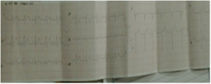
(Photo(1
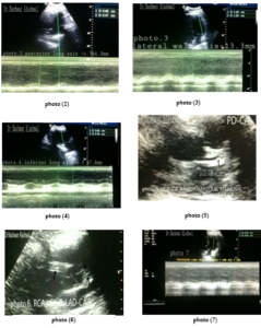
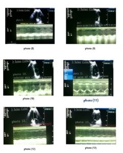
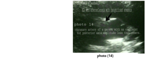
The upper photos: (1) Shown ECG of the patient with non- specific limbs leads changes in leads III, a VL & V1. And photos: (2, 3 and 4) represents the long axes changes in the patient’s heart; posterior axis has variable amplitudes 4.7mm and 6.8mm, lateral axis has 13.3mm while inferior axis has also variable amplitudes= 6.5 and 7.8mm. The lower photo (5): shown the left circumflex coronary artery atheromatous stenosis (dark arrow) and photo: (6): shown three arteries the RCA, LCX and LAD-CA (dark arrows), while the photos : ( 7 & 8): shown the M- mode echo improvements of posterior and inferior axes amplitudes after therapy, and photo No. (9, 10, & 11 )are for long axes amplitudes of person with no complaint ;normal person 2 : shown values above 10 mm with posterior one has 10.5 mm it is less than the other normal 13.3 mm while ( photo 12 & 13 ) are for the patient with ischemic dilated cardiomyopathy shown value of 6.4 mm each one, and the photo (14). Is for right coronary arteries of the normal person 2: shown mild wall atherosclerosis with insignificant lumen stenosis and its walls boundaries are visibly seen.
References:
- Muhammad Ajmal Malik et al., Chest Pain as a presenting complaint in AMI, Pak J Med Sci 2013: 29(2).
- Kevin Channer, Francis Morris, ABC of clinical electrocardiography Myocardial ischemia, BMJ volume:27 April 2002: 324
- Fedorowski & Redden : Stress Testing : Hospital Physician December: 2000: pp. 25–30.
- Marek Krzanowski, Wojciech Bodzo and Pawe Petkow Dimitrow, Imaging of all three coronary arteries by transthoracic echocardiography. an illustrated guide, Cardiovascular Ultrasound, BioMed Central, 2003.
- V. Anjaneyulu, Coronary artery imaging by transthoracic echocardiography: Journal of the Indian Academy of Echocardiography & Cardiovascular Imaging : January – April 2017: 1(1).
- Feigenbaum’s Echocardiography, Evaluation of Systolic and Diastolic Functions of the Left Ventricle, Sixth Edition, by Lippincott Williams & Wilkins: 2005: 6.edit. 140-141.
فحص الشرايين القلبية التاجية بجهاز موجات فوق الصوتية عبر الصدر لمريض بضيق الصدر وألم الظهر،
في عدن- اليمن